

The Enteric Nervous System: Your Gut's Own Brain
The enteric nervous system (ENS) is the intrinsic nervous system of the gastrointestinal tract — a vast, semi-autonomous network embedded in the gut wall that controls digestion, secretion, local blood flow, and mucosal immune responses largely on its own, without waiting for instructions from the brain.
A few things worth knowing right away:
- The ENS contains over 100 million neurons — more than the spinal cord — organized into two primary plexuses that run continuously from esophagus to rectum.
- It can generate complex motor patterns like peristalsis entirely independently of the central nervous system (CNS), earning its nickname “the second brain.”
- The ENS communicates bidirectionally with the brain via the vagus nerve and spinal pathways, meaning gut states influence mood and cognition just as brain states influence digestion.
- It interacts closely with the intestinal microbiome, enteroendocrine cells, and mucosal immune cells — making it a central hub in gut health, not just a digestive relay.
What follows covers ENS anatomy, its cellular architecture, how it develops, what it does, how it talks to the brain and microbiome, what goes wrong when it fails, and how everyday habits support it.
Key Takeaways
The enteric nervous system is a semi-autonomous neural network embedded in the gut wall, capable of controlling digestion independently while remaining in constant dialogue with the brain, microbiome, and immune system.
| Point | Details |
|---|---|
| ENS definition | The intrinsic nervous system of the gut, controlling motility, secretion, blood flow, and immune interactions autonomously. |
| Two plexuses | The myenteric plexus drives motility; the submucosal plexus manages secretion, absorption, and luminal sensing. |
| Neuron scale | The ENS contains over 100 million neurons and uses more than 30 neurotransmitters, rivaling the spinal cord in complexity. |
| ENS disorders | Hirschsprung disease, Chagas disease, diabetic gastroparesis, and IBS all involve ENS dysfunction at different levels of severity. |
| Yakonow at the table | Yakonow yacon syrup provides prebiotic FOS fiber with a glycemic index of 1, a gentle daily detail that supports the gut environment your ENS depends on. |
Table of Contents
- What is the enteric nervous system, and where does it live?
- How the ENS is built: neurons, glia, and the molecules they use
- Where does the ENS come from? Development and congenital links
- What does the ENS actually do? Core functions and the peristaltic reflex
- How the ENS talks to the brain: the gut–brain axis
- The ENS, the microbiome, and the immune system
- What happens when the ENS fails: disorders and symptoms
- How clinicians and researchers assess ENS function
- Practical habits that support a healthy gut environment
- A warm thought from the table
- Yakonow: a small detail that makes mornings better
- Sources
What is the enteric nervous system, and where does it live?
The ENS sits entirely within the wall of the gastrointestinal tract, organized into two concentric networks of ganglia and nerve fibers that span the entire length of the gut. According to NCBI Bookshelf’s neuroscience reference, these two networks are the myenteric (Auerbach’s) plexus and the submucosal (Meissner’s) plexus, and they have distinct locations and distinct jobs.
The myenteric plexus
The myenteric plexus sits between the longitudinal and circular muscle layers of the gut wall. Its primary job is motor control: it coordinates the contraction and relaxation patterns that move contents through the gut, including peristalsis and segmentation. Damage or absence of myenteric ganglia produces severe motility failure.
The submucosal plexus
The submucosal plexus lies in the connective tissue layer between the circular muscle and the mucosa. It monitors the chemical environment of the gut lumen, regulates glandular secretion and fluid absorption, and modulates local blood flow. Think of it as the gut’s sensory and secretory manager, while the myenteric plexus handles the mechanical work.
Regional variation along the gut
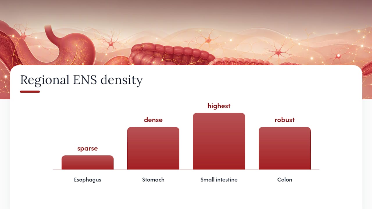
The ENS is not uniform from top to bottom. The esophagus has relatively sparse ganglia; the stomach has a dense myenteric network that coordinates gastric emptying; the small intestine carries the highest density of both plexuses, reflecting its role in nutrient absorption; and the colon has a robust myenteric plexus tuned for slow, mass-movement contractions. This regional specialization means ENS dysfunction can produce very different symptoms depending on which segment is affected.
| Plexus | Location in gut wall | Primary functions |
|---|---|---|
| Myenteric (Auerbach’s) | Between longitudinal and circular muscle layers | Motility coordination, peristalsis, segmentation |
| Submucosal (Meissner’s) | Between circular muscle and mucosa | Secretion, absorption, blood flow, luminal sensing |
How the ENS is built: neurons, glia, and the molecules they use
The cellular architecture of the ENS is far more complex than a simple relay system. StatPearls on the autonomic nervous system notes that the ENS uses more than 30 neurotransmitters — a chemical diversity that rivals the brain itself.
Principal neuron classes
- Intrinsic primary afferent neurons (IPANs): The ENS’s own sensory neurons. They detect mechanical distension and chemical changes in the lumen, then trigger local reflex circuits without involving the CNS.
- Interneurons: Process and relay signals between sensory and motor neurons within the plexus. Ascending interneurons drive excitation orally; descending interneurons drive inhibition anally — the directional logic behind peristalsis.
- Excitatory motor neurons: Release acetylcholine and substance P to contract circular muscle above a bolus.
- Inhibitory motor neurons: Release nitric oxide (NO) and vasoactive intestinal peptide (VIP) to relax circular muscle below a bolus, allowing forward movement.
Enteric glial cells
Enteric glial cells (EGCs) outnumber enteric neurons and are not passive scaffolding. Research published in PMC shows that EGCs actively support neuronal survival, regulate gut barrier integrity, and participate in local immune responses. When EGC function is disrupted, gut permeability and inflammation can follow.
Key neurotransmitters and modulators
| Neurotransmitter | Primary action in the ENS |
|---|---|
| Acetylcholine (ACh) | Excites smooth muscle; drives secretion |
| Nitric oxide (NO) | Relaxes smooth muscle; inhibitory motor neuron signal |
| Vasoactive intestinal peptide (VIP) | Inhibits muscle; promotes secretion and vasodilation |
| Substance P | Excites muscle; involved in pain signaling |
| Serotonin (5-HT) | Initiates peristaltic reflex; modulates gut sensation |
The ENS’s chemical complexity — those 30-plus transmitters — means that drugs targeting one pathway often produce off-target gut effects, which is why so many medications list nausea or constipation as side effects.
Where does the ENS come from? Development and congenital links
The ENS does not arise from the gut itself. Its neurons and glia originate from neural crest cells, a transient, migratory population that forms along the dorsal neural tube during embryonic development.
The developmental sequence runs roughly like this:
- Vagal neural crest cells (from the level of somites 1–7) migrate into the foregut during early embryogenesis and begin colonizing the gut wall in a rostrocaudal direction.
- Sacral neural crest cells contribute a smaller population to the hindgut from the caudal end.
- As these progenitor cells migrate, they proliferate, differentiate into neurons and glia, and form the ganglionic networks of both plexuses.
- The RET receptor tyrosine kinase signaling pathway is critical for this migration and survival. Loss-of-function mutations in RET are the most common genetic cause of failed colonization.
When neural crest migration fails to reach the distal colon, the result is Hirschsprung disease: a segment of aganglionic bowel that cannot relax, causing functional obstruction. Severity depends on how much of the colon lacks ganglia. Surgical removal of the aganglionic segment is the standard treatment, and outcomes depend heavily on how much functional ENS tissue remains.
Hirschsprung is the clearest demonstration that the ENS is not optional. A gut without its intrinsic nervous system cannot move contents forward on its own.
What does the ENS actually do? Core functions and the peristaltic reflex
The ENS’s primary role as local regulator of digestive and defensive functions plays out through four main outputs: motility, secretion, blood flow, and mucosal defense.
The peristaltic reflex, step by step
Peristalsis is the ENS’s signature achievement, and it runs as a local reflex circuit:
- A food bolus distends the gut wall.
- IPANs detect the stretch and chemical signals from the mucosa.
- Ascending interneurons activate excitatory motor neurons above the bolus, contracting circular muscle to push contents forward.
- Descending interneurons activate inhibitory motor neurons below the bolus, relaxing circular muscle to receive contents.
- The bolus moves aborally. The reflex resets and repeats.
Ex vivo gut segments produce this coordinated peristaltic activity even after all extrinsic nerves are severed — direct evidence that the ENS generates the pattern itself. You can read more about what this means for everyday digestion in this guide to gut motility and digestive health.
Other ENS-controlled functions
- Secretion and absorption: The submucosal plexus regulates chloride secretion into the lumen and coordinates fluid absorption, keeping the gut environment hospitable for digestion.
- Local blood flow: ENS neurons release VIP and other vasodilators to increase mucosal blood flow during active digestion.
- Mucosal defense: ENS circuits interact with mast cells and macrophages to modulate inflammatory responses at the epithelial surface.
Autonomy versus CNS modulation
The ENS handles most of its work locally. The sympathetic nervous system generally suppresses ENS activity (slowing motility, reducing secretion) during stress. The parasympathetic system, primarily via the vagus nerve, tends to enhance digestive activity. But these are modulatory inputs, not commands. The ENS runs the show; the brain adjusts the volume.
How the ENS talks to the brain: the gut–brain axis
The gut–brain axis is bidirectional, and the ENS sits at the gut end of that conversation. Signals travel in both directions through distinct anatomical routes.
Ascending (gut to brain): Vagal afferents carry mechanosensory and chemosensory information from the gut to the brainstem nucleus tractus solitarius. Spinal afferents carry pain and discomfort signals to the dorsal horn and then to higher cortical areas. Enteroendocrine cells release peptides (like cholecystokinin and GLP-1) that act on vagal terminals.
Descending (brain to gut): The vagus nerve’s efferent fibers synapse on ENS ganglia, modulating excitability. Sympathetic fibers from prevertebral ganglia inhibit ENS activity during the stress response.
The functional consequences are real and observable. Acute psychological stress can accelerate colonic transit in some people and slow gastric emptying in others, depending on which arm of the autonomic system dominates. The cephalic phase of digestion — the anticipatory secretion of gastric acid and digestive enzymes triggered by the sight and smell of food — is a clean example of descending brain-to-gut signaling that prepares the ENS before a meal even arrives.
What remains an active research area is the precise mechanistic link between chronic psychological states and structural ENS changes. The correlation between stress, anxiety, and gut symptoms is well documented; the causal chain at the cellular level is still being mapped.
The ENS, the microbiome, and the immune system
The ENS does not operate in a sterile environment. It is in constant dialogue with trillions of gut microbes, a specialized population of enteroendocrine cells, and resident immune cells — and disruptions in this cross-talk may contribute to gastrointestinal disease.
Microbe-derived signals that reach ENS neurons
- Short-chain fatty acids (SCFAs): Produced by bacterial fermentation of dietary fiber, SCFAs like butyrate and propionate act on enteroendocrine cells and directly on ENS neurons, influencing motility and secretion.
- Secondary bile acids: Microbially modified bile acids activate receptors on enteroendocrine cells and ENS terminals, modulating colonic transit.
- Bacterial metabolites and neurotransmitter precursors: Some gut bacteria produce or consume serotonin precursors, influencing the pool available for ENS signaling.
Enteroendocrine cell contributions
Enteroendocrine cells (EECs) are scattered throughout the gut epithelium and act as chemosensors. When they detect nutrients, bile acids, or microbial metabolites, they release serotonin, GLP-1, peptide YY, and other signals that activate ENS neurons within milliseconds. This is the gut’s fastest communication channel from lumen to nerve.
ENS and immune cells
ENS neurons and EGCs interact directly with macrophages and mast cells in the gut wall. Mast cells, in particular, can release histamine and other mediators that sensitize ENS sensory neurons, contributing to the visceral hypersensitivity seen in conditions like irritable bowel syndrome. Diet, especially fiber-rich patterns, influences the microbiome and hence ENS signaling — though most mechanistic evidence comes from animal models, and translating specific microbial interventions to human ENS outcomes remains an open question.
Pro Tip: The ENS–microbiome connection is real but nuanced. Avoid interpreting animal-model findings as direct prescriptions for human gut health; the field is moving fast, and human clinical evidence is still catching up.
What happens when the ENS fails: disorders and symptoms
ENS dysfunction spans a wide clinical spectrum, from congenital absence of ganglia to acquired neuropathies and functional disorders. Early enteric neuropathies often present like functional disorders such as IBS, which is why diagnosis can be delayed.
Primary enteric neuropathies
- Hirschsprung disease: Congenital aganglionosis of the distal colon due to failed neural crest migration. Presents as neonatal intestinal obstruction; treated surgically.
- Chagas disease: Infection with Trypanosoma cruzi destroys ENS ganglia, particularly in the esophagus and colon, causing megaesophagus and megacolon.
- Diabetic autonomic neuropathy: Chronic hyperglycemia damages ENS neurons, producing gastroparesis (delayed gastric emptying), constipation, or diarrhea.
- Intestinal pseudo-obstruction (Ogilvie syndrome / chronic intestinal pseudo-obstruction): Severe motility failure without mechanical blockage, often reflecting ENS or extrinsic nerve dysfunction.
Symptom patterns
When ENS circuits are damaged, the gut loses its ability to coordinate movement and secretion. The resulting symptoms depend on location and severity:
- Chronic constipation or complete failure of colonic transit
- Severe nausea and vomiting from gastroparesis
- Abdominal distension and pain from pseudo-obstruction
- Unexplained diarrhea from secretory dysregulation
Disorders of gut–brain interaction
Conditions like irritable bowel syndrome (IBS) and inflammatory bowel disease (IBD) involve ENS changes even when frank neuropathy is absent. In IBS, altered ENS excitability and mast cell sensitization produce visceral hypersensitivity. In IBD, chronic inflammation remodels ENS circuitry, contributing to motility disturbance even during remission.
The ENS contains over 100 million neurons — a scale that explains both its remarkable capacity and the breadth of symptoms that emerge when that network is disrupted. Management of ENS-related disorders typically involves specialist referral (gastroenterology or neurogastroenterology), motility testing, and a combination of medical and, where appropriate, surgical approaches.
How clinicians and researchers assess ENS function
Evaluating the ENS in a living patient is not straightforward: the network is embedded in the gut wall, and most of its activity is invisible from the outside. Clinical tools measure the outputs of ENS function; research tools probe the circuits directly.
Clinical assessment tools
- High-resolution manometry (esophageal, antroduodenal, colonic): Measures pressure patterns generated by ENS-driven muscle contractions; identifies motility disorders and their anatomical location.
- Transit studies (gastric emptying scintigraphy, wireless motility capsule): Quantify how fast contents move through specific gut segments; useful for diagnosing gastroparesis and slow-transit constipation.
- Full-thickness biopsy: The only clinical method that directly samples ENS ganglia; used to confirm Hirschsprung disease and diagnose enteric neuropathies. Requires surgical access.
- Endoscopic mucosal biopsy: Accessible but limited — it reaches only the mucosa, not the myenteric plexus, so it cannot diagnose most enteric neuropathies directly.
Research methods
- Electrophysiology (patch clamp, extracellular recording): Records electrical activity of individual ENS neurons in isolated gut preparations; the gold standard for understanding neuron subtypes and synaptic properties.
- Optogenetics: Allows selective activation or silencing of specific neurochemical classes of ENS neurons using light; single-cell sequencing and optogenetics have clarified ENS cell subtypes and opened new avenues for targeted therapy research.
- Single-cell RNA sequencing: Maps gene expression profiles of individual ENS neurons and glia, revealing far more cell subtypes than classical histology could distinguish.
- Ex vivo motility assays: Isolated gut segments are mounted in organ baths and filmed; software tracks contractile patterns to assess ENS circuit function without anesthesia confounds.
The gap between research and clinic is real. Optogenetics and single-cell sequencing are transforming the understanding of ENS biology in the lab, but translating those insights into clinical diagnostics or therapies is still years away for most applications.
Practical habits that support a healthy gut environment
The ENS is shaped by what surrounds it — and the gut environment is something everyday choices genuinely influence. The evidence base here is strongest for dietary patterns and weakest for specific supplements, so the guidance below stays at the pattern level.
Fiber-rich diets and microbiome diversity are linked to ENS signaling through the SCFA pathway described earlier. Practically, that translates to a few consistent habits:
- Eat varied fiber sources daily. Vegetables, legumes, whole grains, and fruits each feed different microbial populations, broadening the range of metabolites that reach ENS neurons. For family-friendly ideas, the gut microbiome diet staples guide is a good starting point.
- Include fermented foods as tolerated. Yogurt, kefir, kimchi, and sauerkraut introduce live cultures that can shift microbial composition; individual tolerance varies, so start small.
- Keep meal timing regular. The ENS has its own circadian rhythm, and consistent meal times help synchronize motility patterns with the body’s internal clock.
- Limit excess alcohol. Chronic alcohol use disrupts ENS neuron populations and increases gut permeability; moderate intake is far less damaging than habitual heavy use.
- Prioritize sleep and stress management. Chronic stress activates sympathetic pathways that suppress ENS activity; sleep deprivation compounds this. Neither is a trivial lifestyle variable for gut function.
- Include prebiotic fiber sources. Fructooligosaccharides (FOS) and inulin selectively feed beneficial bacteria; FOS fiber and its role in digestion is worth reading if you want the mechanism explained simply.
Pro Tip: If you experience persistent abdominal pain, significant changes in bowel habits, or symptoms that are worsening over time, these habits are not a substitute for evaluation by a gastroenterologist. General lifestyle support and clinical care are not the same thing.
A warm thought from the table
Reading about the ENS, I keep coming back to something simple: the gut is not a passive pipe. It is an intelligent, responsive network that has been quietly coordinating every meal your family has ever shared. That slow Saturday morning with pancakes on the griddle, syrup pooling at the edges, kids still in pajamas — the ENS is already at work before the first bite, primed by the smell of food and the anticipation of gathering.
That is why the small details of the table matter more than we usually give them credit for. What we drizzle, what we pour, what we set out — it all feeds into a system that is paying close attention. Yakonow yacon syrup fits into those mornings as a quiet, considered choice: naturally sweet from the yacon root, with prebiotic FOS fiber that the gut’s microbial community actually uses, and a glycemic index of just 1. Not a supplement, not a health intervention, just a better-tasting detail that makes the moment a little more nourishing.
The art of gathering is also the art of feeding well, without making it complicated.
Yakonow: a small detail that makes mornings better

The science of the ENS points to one practical truth: what you eat consistently shapes the gut environment that your enteric nervous system operates in. Fiber, variety, and gentle prebiotic support matter — not as a regimen, but as a rhythm.
Yakonow yacon syrup is one of the simplest ways to bring prebiotic FOS fiber to the family table without changing the ritual. The glycemic index of 1 means it sits gently on the table for everyone — kids, parents, and guests alike.
Start with the single 6oz bottle for your next slow morning, or grab the pack of 2 so you never run short on a Sunday.
Sources
These are the primary references behind this article, selected for authority and accessibility. They are starting points for deeper reading, not clinical guidance.
- Anatomy, Autonomic Nervous System - StatPearls - NCBI Bookshelf
- Review on ENS interactions with immunity and microbiome - PMC article
- The enteric nervous system | Physiological Reviews